Locations & Hours
- Locations Overview
- DeLand
- New Smyrna Beach
- DeBary
- Port Orange
- Palm Coast
We have 5 locations conveniently located throughout Volusia & Flagler County, Florida.
Please click on a location above or on the map below to see each location's office hours, a map, and more...
DeLand
840 North Stone Street
DeLand, FL 32720
Phone: 386.734.1766

Dr. Jeff Timko

Dr. Charles Heacock

Dr. J Ryan Timko
Hours:
Monday: 7:30-4:30
Tuesday: 8:00-5:30
Wednesday: 7:30-4:30
Thursday: 8:00-6:00
Friday: 7:30-3:00
 
New Smyrna Beach
524 Canal Street
New Smyrna Beach,
FL 32168
Phone: 386.423.5190

Dr. Phillip L.Stephens
Dr. J Ryan Timko
Hours:
Monday: 9:00-5:30
Tuesday: 9:00-5:30
Wednesday: 8:00-5:30
Thursday: 10:00-7:00
Friday: 9:00-12:00
DeBary
2836 Enterprise Rd., Ste 3
DeBary, FL 32713
Phone: 386.668.8885

Dr. Dustin Ramey

Dr. Elizabeth Kester Ramey
Hours:
Mon - Wed: 8:00-5:00
Thursday: 9:00-6:00
Friday: 8:00-12:30
 
Port Orange
5820 S. Williamson Blvd.
Suite 6
Port Orange, FL 32128
Phone: 386.767.4449

Dr. Kirsten Wilgers
Hours:
M-Tu-W: 8:30-5:30
Thursday: 9:00-6:30
Friday: 8:30-12:00
Palm Coast
15 Cypress Branch Way
Suite 206
Palm Coast, FL 32137
Phone: 386.445.1880
Hours:
M-W: 9:00-5:00
Thursday: 9:00-6:00
Friday: 9:00-1:00
Patient Forms
For your convenience, we have provided forms to download & complete before your...
Read more...
NSB Office Moving!
Beginning May 24th, 2013, the New Smyrna Beach Location will be relocating just down the stre...
Read more...
For Physicians
Our Doctors are dedicated to developing mutually beneficial partnerships with area physicians...
Read more...
|

|
|
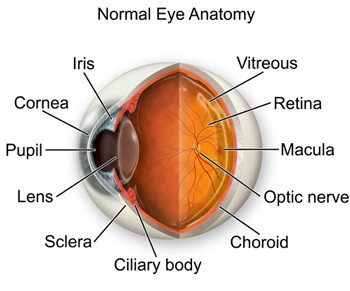
| CORNEA: |
Transparent front segment of the eye that covers iris, pupil, and anterior chamber, and provides most of an eye's optical power. |
| PUPIL: |
Variable-sized, circular opening in center of iris; it appears as a black circle and it regulates the amount of light that enters the eye. |
| IRIS: |
Pigmented tissue lying behind cornea that (1) gives color to the eye, and (2) controls amount of light entering the eye by varying size of black pupillary opening; separates the anterior chamber from the posterior chamber. |
| LENS: |
Natural lens of eye; transparent intraocular tissue that helps bring rays of light to focus on the retina. |
| RETINA: |
Part of the eye that converts images into electrical impulses sent along the optic nerve for transmission back to the brain. Consists ofmany named layers that include rods and cones. |
| MACULA: |
Small, specialized central area of the retina responsible for acute central vision. |
| VITREOUS: |
Transparent, colorless, gelatinous mass; fills rear two-thirds of the interior of the eyeball, between the lens and the retina. |
| OPTIC NERVE: |
Largest sensory nerve of the eye; carries impulses for sight from retina to brain. |
| SCLERA: |
The white of the eye; a protective fibrous outer layer covers all of the eyeball except for the part covered by the cornea. |
| CILIARY BODY: |
A muscular ring under the surface of the eyeball; helps the eye focus by changing the len’s shape and also produces aqueous humor. |
| CHOROID: |
The vascular layer between the sclera and the retina; the blood vessels in the choroid help provide oxygen and nutrients to the eye. |
|
|